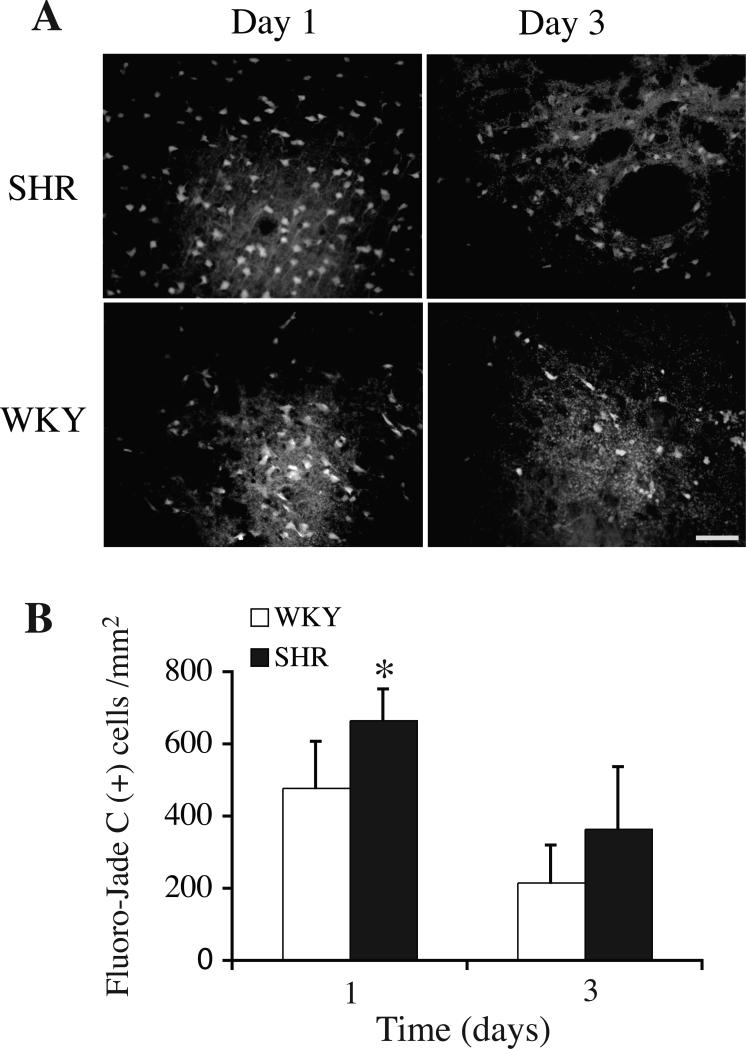
Figure 3.
(A) Fluoro-Jade C staining showing degenerating neurons in the ipsilateral basal ganglia of SHR and WKY rats one and three days after ICH. Bar=50 μm. (B) Bar graph showing numbers of Fluoro–Jade C staining positive cells around the hematoma. Values are expressed as mean±SD. *p<0.05 vs. WKY.

