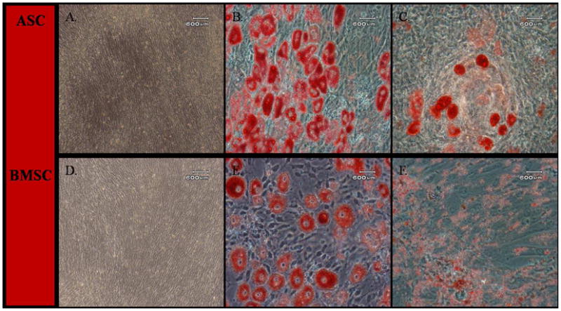
Fig. 3.

Light photomicrographs of canine adipose tissue-derived (ASCs; A–C) and bone marrow-derived stromal cells (BMSCs; D–F) following culture in stromal medium (A, D) or after adipogenic induction for P3 (B, E) and P6 (C, F). The ASCs (A) and BMSCs (D) cultured in stromal medium did not undergo any morphological changes. Following adipogenic induction, P3 and P6 ASCs and BMSCs formed adipocytes with intracellular lipid vacuoles confirmed by Oil Red O staining. P6 ASCs and BMSCs had less lipid accumulation than P3 cells. Scale bar = 600 μm.
