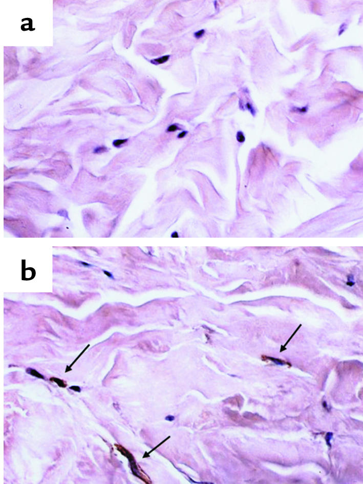
Figure 5.

Expression levels of Smad7 in normal and scleroderma dermal tissue. Paraffin sections of normal dermal tissue (a) and scleroderma dermal tissue (b) were subjected to immunohistochemical analysis with anti-Smad7 Ab, as described in Methods. The immunoreactivity for Smad7 is indicated by arrows.
