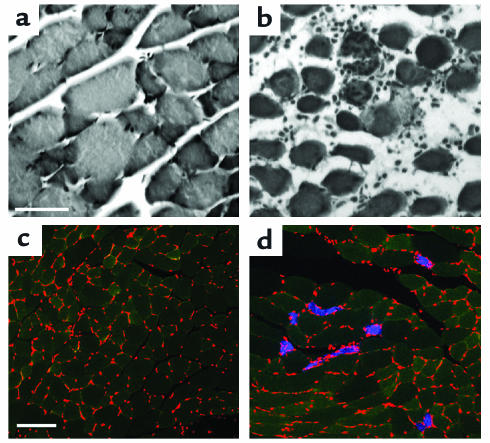
Figure 3.
Histological alterations in Ncx3–/– muscles. H&E-stained sections from 3-month-old Ncx3+/+ (a) and Ncx3–/– (b) gastrocnemius muscle: focus of fiber necrosis and cellular infiltrate in Ncx3–/– mice. Scale bar: 50 μm. Localization of EBD in cryosections of gastrocnemius muscles: 3-month-old EBD-injected Ncx3+/+ (c) and Ncx3–/– (d) mice were examined after 12–24 hours. Red stained structures are nuclei, green stained areas are cytosol, and blue stained areas are EBD-positive fibers. Scale bar: 100 μm.

