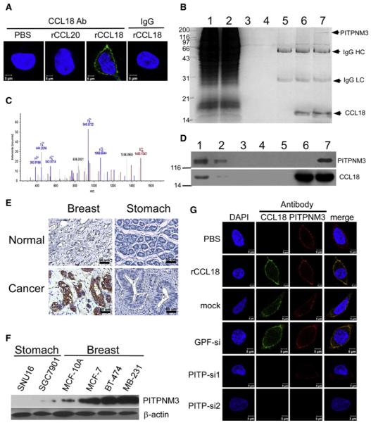
Figure 4. CCL18 Binds to PITPNM3 on Breast Cancer Cell Membrane.
(A) Confocal microscopy for MDA-MB-231 cells treated with PBS, rCCL20, or rCCL18 at 20 ng/ml at 4°C, then fixed and stained with an Alexa 488-labeled anti-CCL18 antibody. An isotype-matched IgG (IgG) was used as a control, and cell nuclei were counterstained with DAPI.
(B) Immunoprecipitation of the membrane extracts (ME) from rCCL18-treated MDA-MB-231 cells with anti-CCL18 antibody. Lane 1 shows input of ME (5%), lane 2 flowthrough (5%), lane 3 ME from the PBS-treated cells incubated with uncoupled protein A/G beads, lane 4 ME from rCCL18-treated cells incubated with uncoupled beads, lane 5 ME from PBS-treated cells incubated with antibody-coupled beads, lane 6 SDS-treated ME from rCCL18-treated cells incubated with antibody-coupled beads, and lane 7 ME from rCCL18-treated cells incubated with antibody-coupled beads. IgG HC, IgG heavy chain; IgG LC, IgG light chain.
(C) Mass spectra of a representative peptide fragment from the protein band indicated by an arrow in lane 7 of (B).
(D) Western blot validation of mass spectrometric identification using an anti-PITPNM3 antibody (upper) and an anti-CCL18 antibody (lower). All lanes and conditions are described as in (B).
(E) Immunohistochemistry for PITPNM3 expression in breast and gastric carcinomas, as well as normal breast and gastric tissues in paraffin tissue sections.
(F) Western blotting for PITPNM3 expression in gastric cancer cell lines of SGC7901 and SNU16, breast epithelial MCF-10A line, and breast cancer cell lines of MCF-7, BT-474, and MDA-MB-231. β-Actin was used as a loading control.
(G) Confocal microscopy for MDA-MB-231 cells stained with an Alexa 488-labeled anti-CCL18 antibody and a Cy3-labeled anti-PITPNM3 antibody. The cells were treated with PBS (row 1) or recombinant CCL18 (rCCL18) at 20 ng/ml (rows 2–6) for 3 hr at 4°C, and were untransfected (row 2), mock transfected (row 3), transfected with GFP-siRNA (row 4), or either of the two PITPNM3-siRNAs (rows 5 and 6). Cell nuclei were counterstained with DAPI.
See also Figure S4.

