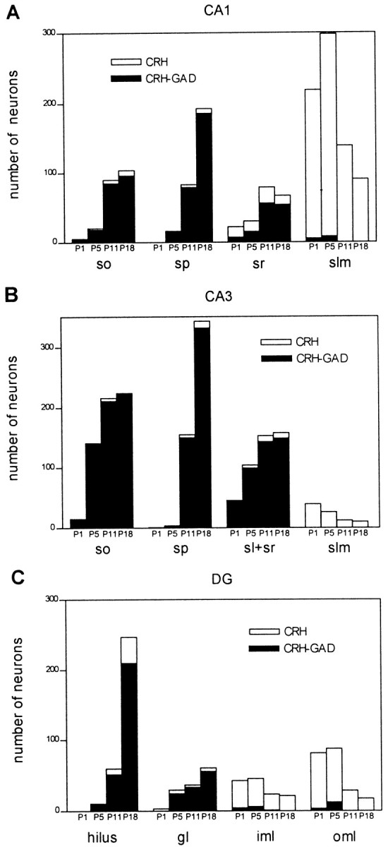
Fig. 5.

Quantitative analysis of GAD67 mRNA expression defines separate hippocampal CRH-expressing neuronal populations, with distinct spatial and age-dependent distribution profiles. Plotting GABAergic CRH-immunoreactive neurons as a proportion of total peptide-expressing cells reveals that the former provide the major contribution to the striking increase of CRH-expressing cells in the principal cell layers between P1 and P18. In contrast, the transient populations in the hippocampal marginal zones [slm and outer molecular layer (oml)] are non-GABAergic. Mixed populations reside in the polymorphic layers, sr, and the inner molecular layer (iml). Quantitative data were derived from eight sections per rat (see Materials and Methods) and two animals per age group.
