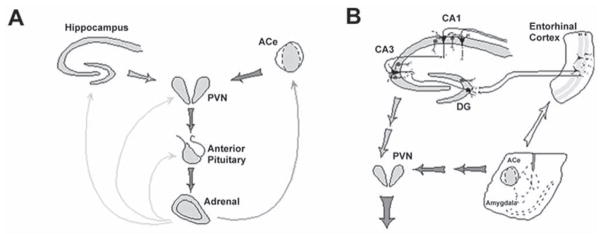
Fig. 1.
The neuroendocrine (A) and limbic (B), stress-activated CRH–ACTH-steroid loops. (A) Stress-conveying signals rapidly activate CRH-expressing neurons of the central nucleus of the amygdala (ACe). Rapid CRH release in the ACe activates CRH-expression in the hypothalamic paraventricular nucleus (PVN) to secrete CRH into the hypothalamo-pituitary portal system, inducing ACTH and glucocorticoid (steroid) secretion from the pituitary and adrenal, respectively. Steroids exert a negative feedback on the production of CRH in the hypothalamus (directly and via the hippocampus), yet activate CRH gene expression in the amygdala, potentially promoting further CRH release and seizure-promoting actions in this region. (B) CRH-expressing GABAergic interneurons (dark cells) in the principal cell layers of the hippocampal CA1, CA3 and the dentate gyrus (DG) are positioned to control excitability of the pyramidal and granule cells, respectively. These neurons may be influenced by stress-evoked release of CRH from the ACe, via connections in the entorhinal cortex. For both panels, thick and thin arrows denote established or putative potentiating and inhibitory actions, respectively. Arrows do not imply mono-synaptic connections. (Modified and published with permission, from Baram and Hatalski [15]).

