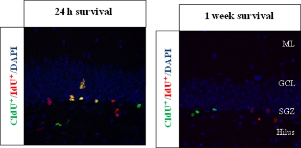
Figure 2.
Representative pictures of CldU+ and IdU+ cells in set-up experiments. The thymidine analogs were injected in the same individuals separated by either 24 h or 1 week; BrdU-equimolecular dosages of CldU and IdU were injected. Animals were then sacrificed 2 h after the last injection. We found that the injection of different thymidine analogs separated by more than 1 day (1 week survival) led to no overlapping of the labeling, while in the 24 h experiment a huge proportion of cells were co-labeled, as expected. The labeling was assessed in the hippocampal dentate gyrus of adult mice. The pictures were registered from 50 μm thick coronal sections, and both are taken from Llorens-Martin et al. (2010). ML, molecular layer; GCL, granule cell layer; SGZ, subgranular zone.

