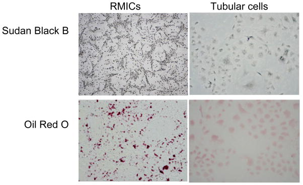
Figure 10. Sudan Black B and Oil Red O staining in RMIC and tubular cells.
The cell shapes were apparently different between these two types of cells. There were numerous positive staining vesicles in RMICs. In contrast, there were few such positive staining vesicles observed in tubular cells. (200 ×)

