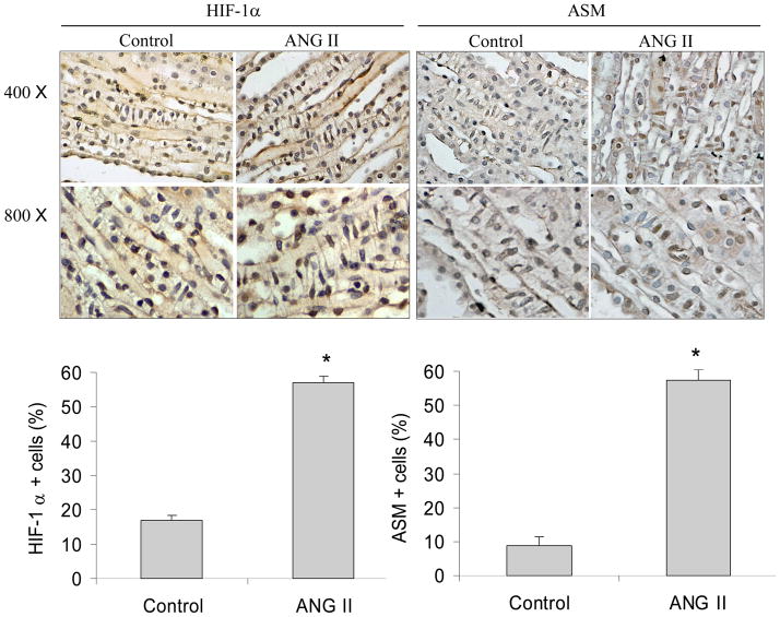
Figure 9. Immunohistochemical staining of HIF-1α and SMA in the renal inner medulla.
Top panels: Representative photomicrographs. Brown color indicates positive staining. Bottom panels: Percentage quantitation of positive cells. * P<0.05 vs. control (n=5). RMICs were identified by their unique morphological features of a ladder-like arrangement with the long axis of the cells perpendicular to the long axis of the papilla.

