
Figure 4.
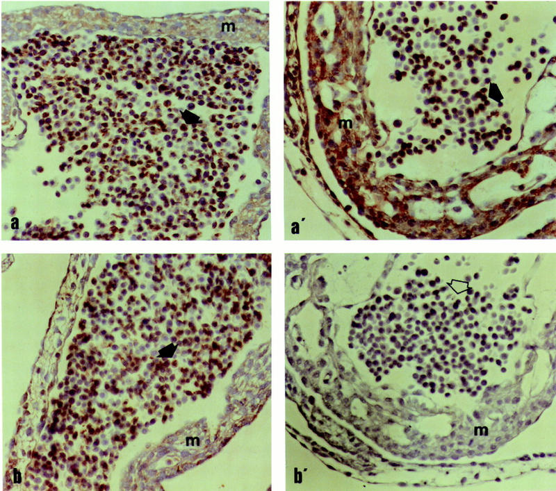
Light microphotographs of the cardiac wall (m = endomyocardium) and blood cells (arrows) in dominant white I/I (a,b) and colored embryos i/i (a′,b′) after incubation with the Ex21 (a,a′) and Ex16/18 (b,b′) antibodies. Note the negative immunolabeling of Ex16/18 in colored embryos (b′, open arrow). ABC-ELITE, magnification 200×.