
Figure 3. Mitochondrial hypothesis and AD.
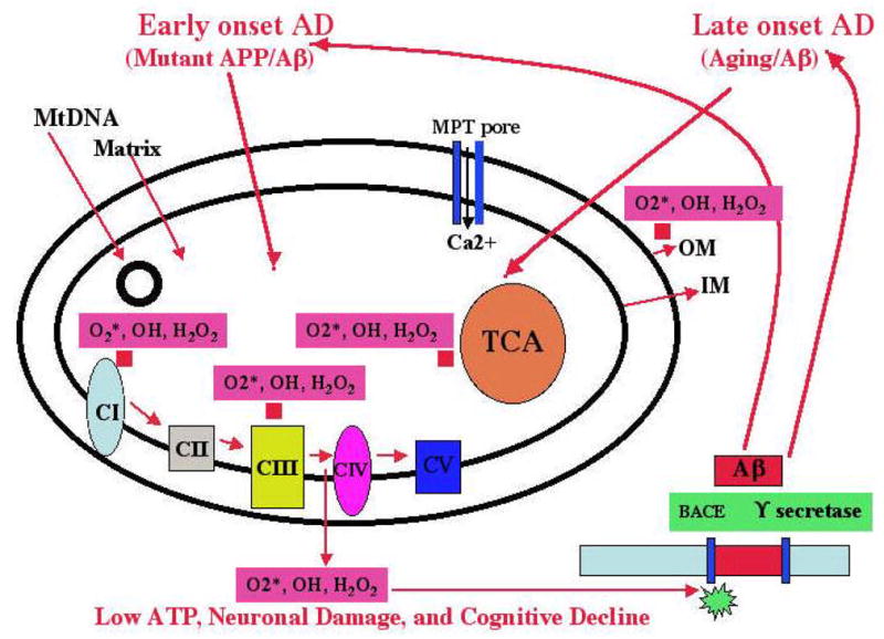
In early-onset AD, mutant APP and soluble Aβ are hypothesized to localize to synaptic mitochondria, leading to the generation of free radicals (O2*, H2O2, OH). The free radicals, in turn, may decrease cytochrome oxidase activity and inhibit cellular ATP. In late-onset AD, the free radicals that are generated due to aging may activate BACE and facilitate the cleavage of the Aβ. The cleaved Aβ may enter mitochondria and induce free radicals (O2*, H2O2, OH), leading to the disruption of the ETC, a decrease in cytochrome oxidase activity, and the inhibition of ATP. This feedback loop may ultimately lead to neuronal damage, to the degeneration of neurons, and to cognitive decline in AD patients.