
Figure 5.

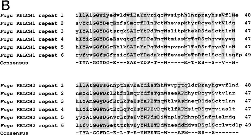
(A) Alignment of Fugu KELCH1 and KELCH2 proteins. The alignment was performed with the GCG programs PILEUP and PRETTY. Residues identical at each position are shown in uppercase letters and shaded. Intron/exon boundaries are shown by closed arrowheads on either site of the alignment. Each repeated segment is boxed and the beginning of each repeat is indicated by its respective number. (B) Alignment of the six repeated segments of Fugu KELCH1 and KELCH2 proteins. The alignments were performed with the GCG programs PILEUP and PRETTY. Amino acid residues at least identical in three repeats are shaded and shown in the consensus sequence. Dashes were introduced at less conserved positions.