Figure 5.
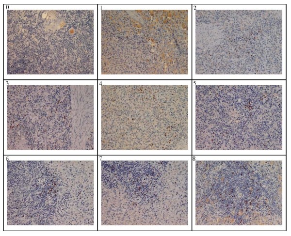
CSFV antigen localization based on WH303-mediated immune-histochemistry for spleen from day 1 to 8. IHC stains for CSFV in spleen of infection pigs euthanized form day 1 to day 8 post infection. The cells positive for viral antigens appeared dark-brown. Picture 0 was the negative control and 1, 2, 3, 4, 5, 6, 7 and 8 were day 1 to day 8 post infection respectively. The CSFV antigen positive cells were distributed in the medulla, cortex, dendritic reticulum and follicular epithelium. Magnification 400×.

