
Figure 2.
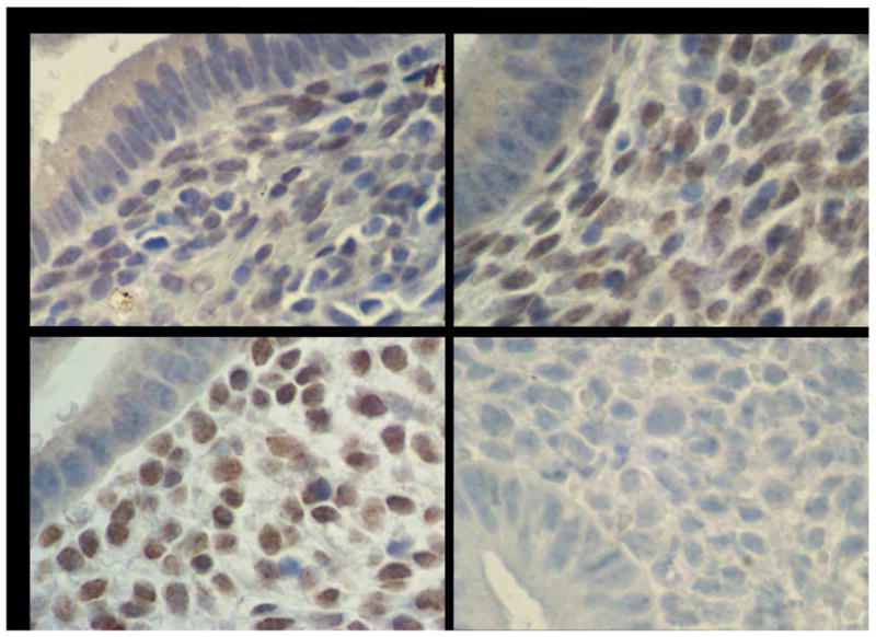
Endometrial HOXA10 Immunohistochemistry
Immunohistochemistry identified HOXA10 protein expression in endometrial glands and stroma. H-SCOREs were determined separately for the glands and stroma. Shown are representative photomicrographs demonstrating HOXA10 expression in endometrium in the setting of: (A) Submucosal myoma, (B) Intramural myoma, (C) No myomas, (D) HOXA10 negative control omitting primary antibody.