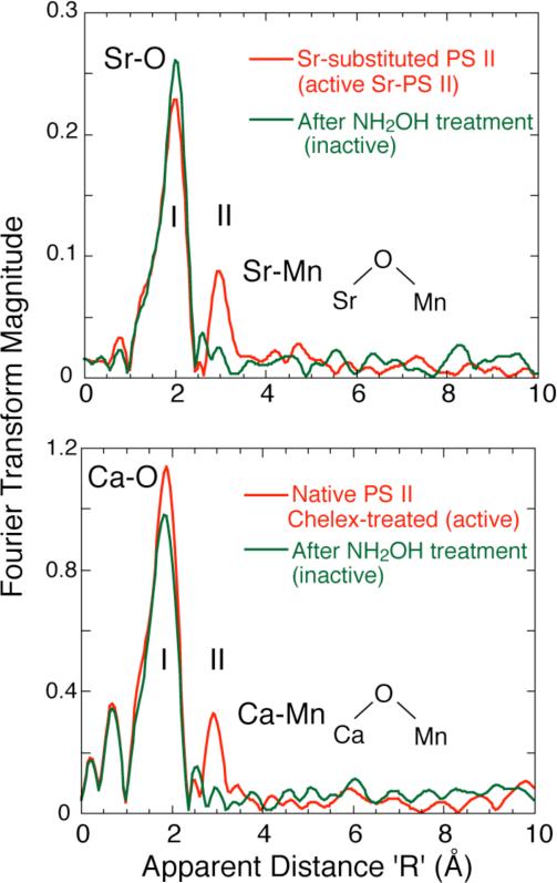
Figure 2.
Top. Fourier transforms of Sr EXAFS for intact and inactive Sr-substituted PS II samples (Chelex-treated). The dominant Fourier Peak I is due to ligating oxygens in the first coordination sphere and is common to both samples. The two Sr-PS II samples differ mainly at the R’ = 3.0 Å region, where the intact samples exhibit Fourier Peak II, which is from Sr-Mn vectors. Disruption of the Mn cluster in NH2OH-treated sample leads to the absence of Peak II. Bottom. Fourier Transform of Ca EXAFS from Chelex-treated, layered samples with 2 Ca/PS II with O2-evolving activity and an S2 EPR multiline signal. The FTs show the presence of a second Fourier peak in the 2Ca/PS II sample that fits to Ca-Mn that is absent in the control sample where the Mn complex has been disrupted with NH2OH.

