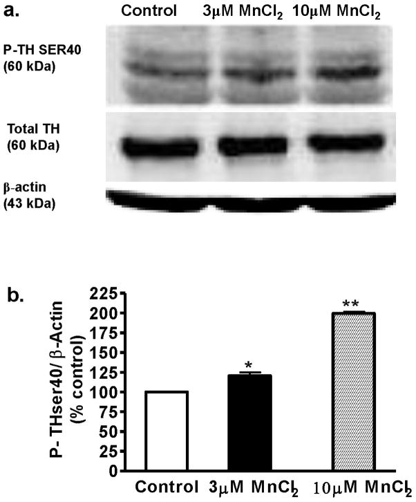
Fig. 2.
Effect of acute Mn treatment on TH phosphorylation level in differentiated N27 cells. Differentiated N27 cells were exposed to 3 or 10 μM MnCl2 for 3 h. Cell extracts were prepared and separated by SDS-polyacrylamide gel electrophoresis and transferred to nitrocellulose membrane. TH antibody (mouse, 1:1000) and phospho-specific antibodies directed against P-TH-Ser40 (rabbit, 1:1000) were used for immunoblotting. To confirm equal protein loading in each lane, the membranes were reprobed with β-actin antibody. The immunoblots were visualized using Amersham’s ECL detection agents. Densitometric analysis of 60 kDa P-TH-Ser40 bands represents the mean ± SEM from three separate experiments (*p < 0.05, **p < 0.01).

