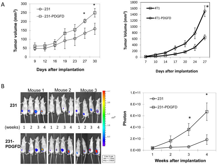
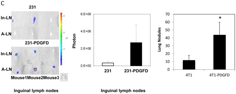
Figure 2.
(A) Parental and PDGF-D transfected MDA-MB-231 and 4T1 cells were injected into the fourth left mammary fat pad of nude mice, tumor size was measured every 3 days (n = 8, P < 0.005). (B) Representative bioluminescence images (BLI) of mice bearing parental (231) and PDGF-D transfected (231-PDGFD) MDA-MB-231 tumors. BLI study started 7 days after tumor implantation and was performed weekly to monitor the development of tumors. Signal intensity is measured as photon flux (photons/second) and coded to a color scale. (C) Thirty days after tumor implantation, mice bearing parental and PDGF-D transfected MDA-MB-231 tumors were sacrificed. Inguinal (In-LN) and axillary (A-LN) lymph nodes were collected for BLI study (n = 8). BLI images were quantified using Living Image software 3.0. P < 0.005. Lungs were collected from mice bearing parental and PDGF-D transfected 4T1 tumors, metastatic nodules were counted under dissecting microscope.


