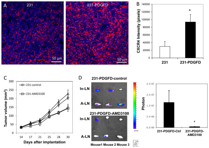
Figure 4.
(A) Immunofluorescent staining of CXCR4 in parental MDA-MB-231 and 231-PDGFD tumor tissues (20x). (B) Quantification of CXCR4 staining intensity by ImageJ. P < 0.01. (C) Fourteen days after orthotopic implantation, mice were randomized to be implanted subcutaneously with a 14-day continuous release osmotic pump (Alzet®, Cupertino, CA) containing saline (control), or AMD3100 (7 mg/kg/day) (n = 8). Tumor size was measured every three days by caliper. Mice were sacrificed on day 30, and (D) inguinal (In-LN) and axillary (A-LN) lymph nodes were collected for BLI study. BLI images were quantified using Living Image software 3.0. P < 0.005.

