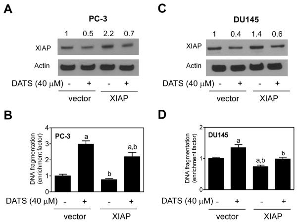
Fig. 4.
Immunoblotting for XIAP protein using lysates from PC-3 (A) and DU145 (C) cells transiently transfected with empty pcDNA3.1 vector or pcDNA3.1 vector encoding for XIAP and treated with DMSO (control) or 40 μmol/L DATS for 16-24 hours. Numbers above the bands represent change in protein level relative to empty vector-transfected cells treated with DMSO. Histone-associated DNA fragment release into the cytosol (a measure of apoptotic cell death) in PC-3 (B) and DU145 (D) cells transiently transfected with empty pcDNA3.1 vector or pcDNA3.1 vector encoding for XIAP and treated with DMSO (control) or 40 μmol/L DATS for 16-24 hours. Columns, mean (n= 3); bars, SD. Significantly different (P < 0.05) compared with empty vector-transfected cells treated with DMSO (a) and empty vector-transfected cells treated with DATS (b) by one-way ANOVA followed by Bonferroni's multiple comparison test. Comparable results were observed in two independent experiments. Representative data from one such experiment are shown.

