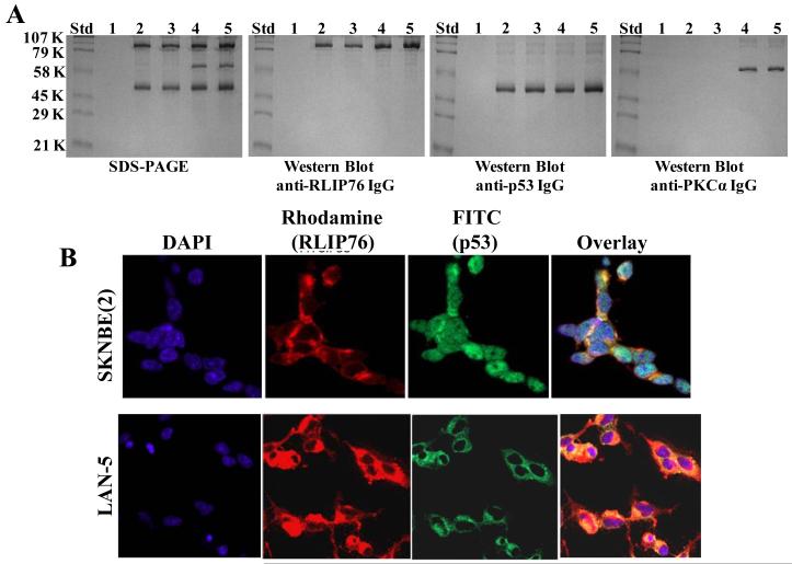
Figure 2.
Interaction of RLIP76 with p53: Purified rec-RLIP76 and p53 proteins (10 μg each), in the absence/presence of equimolar-concentration of PKCα and/or 1 mM ATP were incubated for 30 min at 37 °C and were cross-linked and immuno-precipitated as described in the methods. Cross-linked proteins were resuspended in 100 μl of SDS-PAGE sample-buffer and analyzed by SDS-PAGE, followed by Western-blots against anti-RLIP76 IgG, anti-p53 IgG, and anti-PKCα IgG. Lanes 1 to 5 shows the control with pre-immune IgG (lane 1); immuno-precipitated RLIP76 and p53 (lane 2); RLIP76, p53 and 1 mM ATP (lane 3); RLIP76, p53 and PKCα (lane 4), and RLIP76, p53, PKCα and 1 mM ATP (lane 5), respectively (panel A). Immuno-histochemical localization of RLIP76 and p53 in p53-mutant (SKN-BE2) and p53-normal (LAN-5) neuroblastoma cells-Red: Rhodamine for RLIP76; Green: FITC for p53, yellow overlay indicating co-localization (panel B).

