
Fig. 2. Cell surface Axl expression.
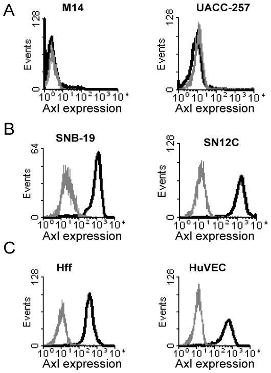
A) Axl surface expression on two NCI-60 cell lines that were poorly transduced with ZEBOV-GP ΔO VSV and displayed low levels of AXL expression in NCI-60 gene array studies. B) Axl surface expression on two NCI-60 cell lines that were highly transduced with ZEBOV-GP ΔO VSV and displayed robust levels of AXL mRNA expression on the gene arrays. C) Axl expression on the surface of two primary human cell populations, human foreskin fibroblasts (Hff) and umbilical cord endothelial cells (HuVEC). Cells stained with normal goat sera are shown in grey histograms, whereas cells stained with goat anti-human Axl antisera are shown in black histograms.