
Fig. 6. Axl is required for post-binding events.
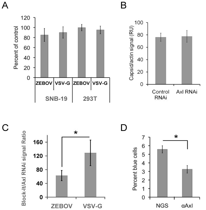
A) Soluble Axl-Fc does not interfere with ZEBOV-GP ΔO pseudotyped VSV transduction. ZEBOV and VSV-G virions were pre-incubated with Axl-Fc (50 μg/mL) and transduced on to Axl dependent SNB-19 cells or the Axl-independent 293T cells. Transduction was evaluated by EGFP expression in the transduced populations at 24 hours. Results are shown as the number of cells transduced in the presence of Axl-Fc divided by the number of transduced cells in the absence of treatment. B) RNAi knock down of Axl had no effect on ZEBOV-GP ΔO pseudovirion binding. Forty-eight hours following transfection of AXL RNAi or an irrelevant RNAi into SNB-19 cells, equivalent quantities of ZEBOV-GP ΔO FIV were incubated with cells for one hour at 4°C. Unbound virus was removed and cells were lysed. Lysates were immunoblotted for FIV capsid and quantitated as described in the Materials and Methods. Shown is average pixel values of FIV capsid on the immunoblot divided by the average pixel values for cellular β-actin from 10 independent experiments. C) ZEBOV-GP ΔO FIV internalization, but not VSV-G FIV is decreased in AXL siRNA-treated cells. SNB-19 cells were transfected with AXL siRNA or an irrelevant control siRNA. At 48 hours, ZEBOV-GP ΔO FIV pseudovirions were bound to the cells for 1 hour at 4°C. Unbound virus was removed and cells were shifted to 37°C for 2 hour. Cells were lysed and immunoblotted for FIV capsid and cellular actin. The capsid signal was normalized for actin levels and data are reported as the ratio of the FIV signal in the Axl knock down cells divided by the FIV signal in the cells transfected with an irrelevant RNA. D) Ability of Axl antisera to block ZEBOV VLP fusion events. ZEBOV-GP ΔO-VLPs containing Src-β-lactamase were transduced into SNB-19 cells in the presence or absence of 1:20 dilution of polyclonal antisera against the ectodomain of Axl or normal goat sera (NGS). Entry of β-lactamase into the cytoplasm of cells was evaluated by flow cytometry following incubation of the cells with the fluorescent β-lactamase substrate CCF2/AM for 2 hours. *p < 0.05.