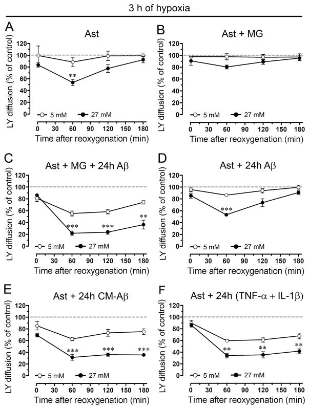
Figure 2. Aβ25-35-treated microglia potentiate the reduction of astroglial coupling induced by hypoxia in high glucose.
(A–F) Averaged data normalized to control (dashed line) of area of LY diffusion in astrocytes alone (Ast) or astrocytes co-cultured for 24 h with microglia (MG) in presence or absence of 10 μM Aβ25-35 (Aβ) and then exposed to 3 h hypoxia in 5 mM (○) or 27 mM (●) glucose followed by several periods of reoxygenation. It is shown the LY diffusion between astrocyte pre-treated for 24 h with 10 μM Aβ25-35 or with conditioned media from microglia exposed for 24 h to 10 μM Aβ25-35 (CM-Aβ) and then subjected to hypoxia/reoxygenation. It is also shown the LY diffusion between astrocytes pre-treated for 24 h with TNF-α and IL-1β (10 pg/ml of each).** p < 0.005, *** p < 0.001, (●) vs (○) at each time point. Each value corresponds to mean ± S.E. of four independent experiments.

