
Figure 5. Aβ25-35-treated microglia induces increase in surface levels of Cx43 after hypoxia.
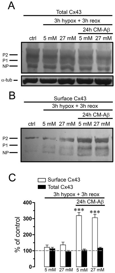
(A and B) Astrocytes cultures were controls or were subjected to 3 h hypoxia in 5 or 27 mM glucose followed by 3 h reoxygenation. Other cultures were pre-incubated for 24 h in CM-Aβ and then subjected to 3 h hypoxia in 5 or 27 mM glucose followed by 3 h reoxygenation. Levels of total Cx43 and surface Cx43 isolated by biotinylation were measured by Western blot analysis. (A) Western blot of total Cx43 present in homogenates. None of the treatments affected quantities of the phosphorylated (P1–P2) and nonphosphorylated (NP) forms of Cx43 (markers on the left). (B) Western blot of surface Cx43 from astrocytes under the same conditions. (C) Quantification of surface and total Cx43 normalized to the control in the treatments mentioned above. *** p < 0.001, compared to control. Each value corresponds to mean ± S.E. of at least 3 independent experiments.