Figure 2.
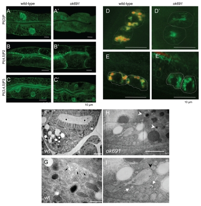
bec-1 mutants display defects in endocytosis. The detection of the phosphoinositides PI3P, PIP2 and PIP3 in the intestines of wild-type (A–C), bec-1(ok691) mutants (A'–C'), animals is shown. Scale bars indicate 10 µm. In wild-type animals (A), PI3P is enriched in the apical membrane with weak basolateral labeling. In bec-1(ok691) (A') mutants very low and diffused expression of PI3P was observed. PIP2 expression is not affected in bec-1(ok691) mutants (B') when compared to wild-type animals (B). The PIP3 reporter labels apical membrane and strong basolateral expression in wild-type animals (C). The localization of PIP3 in bec-1(ok691) animals (C') was similar to that of wild-type animals (C). Micrographs of LMP-1::GFP positive lysosomes in coelomocytes 24 h after the injection of Texas Red BSA (a fluid phase marker for endocytosis) into the body cavity (pseudocoelom). A hatched line demarcates the periphery of the coeolomocytes. The red dye accumulates in lysosomes (LMP-1 positive) localized in the coelomocytes of the wild-type animals (D), but fails to accumulate in the lysosomes (LMP-1 positive) of bec-1(ok691) mutants (D'). Micrographs of LMP-1::GFP positive lysosomes in coelomocytes after 1 h of soaking in Texas Red BSA (a fluid phase marker for endocytosis). The red dye accumulates in lysosomes (green) localized in the coelomocytes of the wild-type animals (E), but fails to accumulate in the lysosomes (green) of bec-1 mutants (E'). Electron microscopy of an N2 wild-type (F and G) and bec-1(ok691) mutant young adults (H and I). (F) Low power electron micrograph of wild-type adult intestine in cross-section, fixed by high pressure freezing. Most of the basal membrane shows few signs of endocytosis, although there is some membrane infolding (black arrow in F) and a few small vesicles, particularly near the lateral membrane border between two intestinal cells on the extreme left edge of the tissue. Higher power view of adult wild-type intestine shows a few examples of membrane infolding (black arrows in G) representing a local zone where endocytosis is active. It is notable that the infolded membranes are rather large, and there are almost no vesicles nearby in the cytoplasm. In contrast, chains of vesicles (white arrows in I) and extensive infolded membranes are seen along the basal pole in the bec-1 mutant (H and I). Thus endocytotic events must be cleared relatively quickly in the wild-type intestine. In bec-1 mutant intestines, defective enlarged endosomes (marked by white arrow head in H) are seen that correspond to abnormal vacuoles of different sizes and contain either membranes or condensed material. (I) is a close up of the region marked in (H) and it shows enlarged vacuoles and a high number of early endosomes that appear to be fusing in some cases (black arrowhead in I). This phenotype is never seen in the intestines of wild-type animals (F or G).

