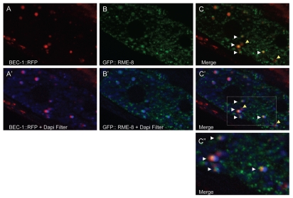
Figure 4.
BEC-1 colocalizes with RME-8. Representative images of intestinally expressed RFP-tagged BEC-1 (red, A, A') and GFP-tagged RME-8 (green, B, B') in wild-type intact living animals. Autofluorescent lysosome-like organelles are shown in blue (+ DAPI filter, D). White arrowheads indicate colocalization of the BEC-1::RFP and RME-8::GFP signals (C), observed as the yellow fluorescence. The blue autofluorescence is also shown with BEC-1::RFP (A') and with GFP::RME-8 (B') and in the merger of both in (C'). The colocalized BEC-1::RFP and GFP::RME-8 signals are found adjacent to the autofluorescent lysosomes which can be visualized with the DAPI filter (C'). A close-up of (C') is shown in (C”). Arrowheads in yellow show BEC-1::RFP that is not associated with RME-8::GFP. Magnification is 630x (A–C').

