
Figure 4.
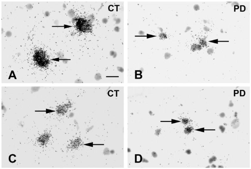
Bright-field photomicrographs illustrating GAD67 mRNA labeling on emulsion radioautographs in prefrontal cortex BA9. Cases are from the HBTRC. Arrows indicate clusters of silver grains over GAD67 mRNA labeled neurons. A=Control. B=PD case. C=Control. D=PD case. Scale bar: 20μm.