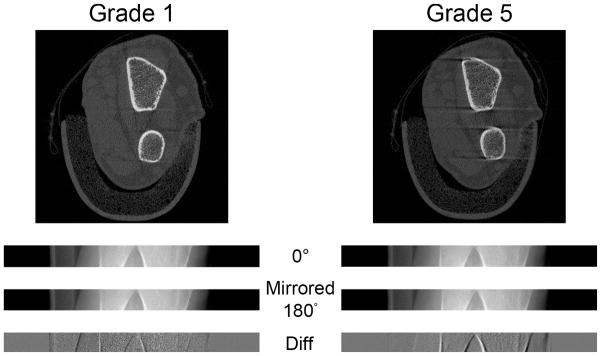
Figure 3.
Comparison of radius scans from a subject with a grade 1 score (left) and a grade 5 score (right). From the top: the reconstructed images, parallelized projection images at 0°, flipped parallelized projection images at 180°, and the difference images of the two. For grade 5, the flipped parallelized projection image at 180° is shifted to the right with respect to the parallelized projection image at 0°. This offset is also apparent in the difference image with more defined edges of the projected bone contour

