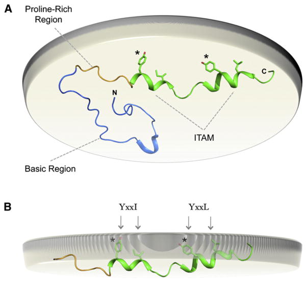
Figure 3. Regulation of Immunoreceptor Signaling through Dynamic Membrane Binding of Cytoplasmic Signaling Sequences.
(A) Solution NMR structure of the membrane-bound mouse CD3ε cytoplasmic tail sequence (PDB ID 2K4F) (Xu et al., 2008). A view from the perspective of the cell interior is shown with the inner leaflet of the plasma membrane bilayer represented by the beige disc. Membrane binding is energetically dominated by the polybasic region (blue) directly C-terminal to the TM domain. The proline-rich region (yellow) and the ITAM (green) follow.
(B) In the membrane-bound structure, the ITAM region adopts a partially helical conformation and inserts the canonical tyrosine and aliphatic residues into the lipid bilayer interior where they are protected from phosphorylation by Src-family kinases.

