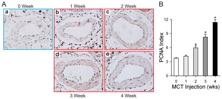
Figure 2. MCT-induced PH is characterized by increased pulmonary artery smooth muscle cell proliferation.
Lung sections from each rat in the different groups were fixed in formalin and embedded in paraffin. PCNA staining was used to indicate pulmonary artery smooth muscle cell proliferation over the time course indicated. Representative sections are shown in (A) and the summarized data in (B). Data are presented as mean ± SD; n=8/group; * P<0.05 vs. 0 week group.

