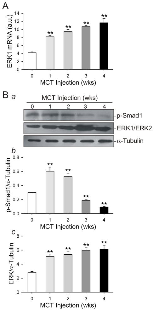
Figure 3. MCT induces an increase in ERK protein levels and a decrease in activated (or phosphorylated) Smad1 protein in whole rat lung.
mRNA and protein were collected from whole lung from rats injected with MCT at the indicated timepoints after injection. The week 0 group was injected with saline, and protein and mRNA was collected the next day. mRNA was used in RT-PCR to quantify ERK1 mRNA levels (A) using the primer listed in Table 1. This signal was normalized against GAPDH mRNA levels. Protein lysate resolved electrophoretically was transferred to a nitrocellulose membrane which was immunoblotted for phosphorylated Smad1 (p-Smad1) and ERK. Representative gels are shown in (Ba) and summarized data in (Bb and Bc). Protein signals were normalized to a-tubulin protein levels. Data are presented in arbitrary units (a.u.) as mean ± SD; n=5/group; ** P<0.01 vs. 0 week control group.

