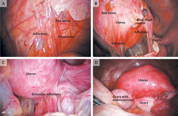
Figure 1. Peritoneal Lesions and an Ovarian Endometrioma Due to Endometriosis.
Panel A shows an endometriotic implant (red lesion), adhesions, and hyperemia in the peritoneum. Panel B shows peritoneal implants, including red and blue–black lesions and adhesions. Panel C shows extensive adhesions distorting the normal pelvic anatomy. Panel D shows an endometrioma adherent to the posterior uterus and distending the ovarian capsule. At surgery, it is difficult to distinguish visually an endometrioma from a cyst of the corpus luteum, a hemorrhagic cyst, or a simple cyst. Although the cyst fluid in endometriomas is thick and dark brown because it contains hemosiderin (hence, the name “chocolate cysts”), this color is not specific to endometriomas. (Images courtesy of Dr. Christopher Herndon, University of California, San Francisco.)

