
Fig. 4.
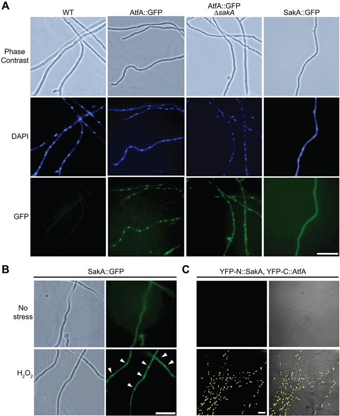
AtfA::GFP shows nuclear localization independently of sakA; SakA accumulates in the nucleus in response to hydrogen peroxide stress, where it interacts with transcription factor AtfA.
A. Conidia from strains CLK43 (WT), TFL3 (AtfA::GFP), CFL9 (ΔsakA; AtfA::GFP) and TFL6 (SakA::GFP) were inoculated on coverslips submerged in liquid supplemented minimal medium, incubated for 12 h at 37°C and then fixed and stained with DAPI.
B. Conidia from strain TFL6 were inoculated as in (A) and then treated with 30 mM H2O2 for 30 min. Arrowheads indicate SakA::GFP localization after stress treatment. Pictures were taken using Epifluorescence with a microscope NIKON Eclipse E600.
C. BiFC analysis of SakA and AtfA. Strain TFL7 expressing YFP-N::SakA and YFP-C::AtfA was grown as in (B), transferred to minimal medium with 100 mM threonine for 3 h to induce the alcA promoter, and exposed to 30 mM H2O2 for 30 min. Yellow fluorescence was detected using a confocal microscope Olympus FV1000.Bar = 20 µm.