
Fig. 8.
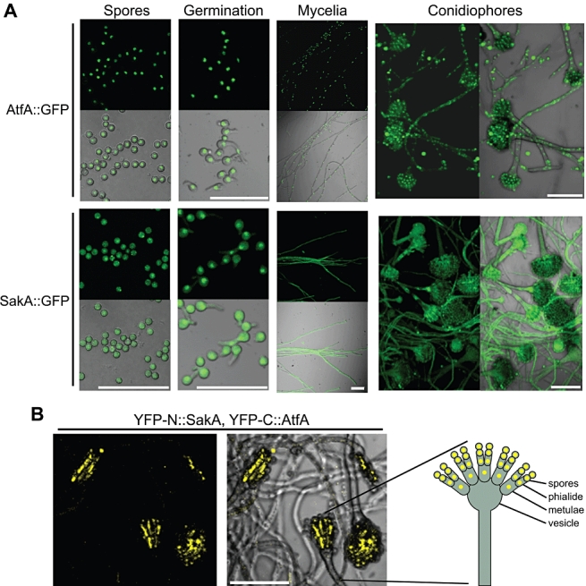
SakA and AtfA localization during growth and development; SakA and AtfA interact during conidiophore development.
A. Spores from strains TFL3 (AtfA::GFP) and TFL6 (SakA::GFP) were inoculated on supplemented MM plates and observed before germination (Spores), 4 h after germination (Germination), after 24 h of growth (Mycelia) and after 48 h of induction of conidiation (Conidiophores).
B. Strain TFL7, expressing YFP-N::SakA and YFP-C::AtfA, was inoculated on a plate containing MM and 100 mM threonine, to induce the alcA promoter, and observed after 48 h. A schematic representation of a conidiophore indicates cell types showing YFP fluorescence. Pictures show Z stacks of confocal images obtained with the confocal microscope Olympus FV1000.Bars = 30 µm.