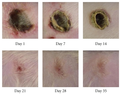
Figure 3.
Typical macroscopic view of pressure ulcer of control group on days 1, 7, 14, 21, 28, and 35. Pressure-loaded portion became necrotic after release of loading (day 1), and crust was formed in the ulcer site with redness of surrounding skin (day 7). Size of ulcer gradually decreased (days 14, 21, and 28), and disappeared (day 35).

