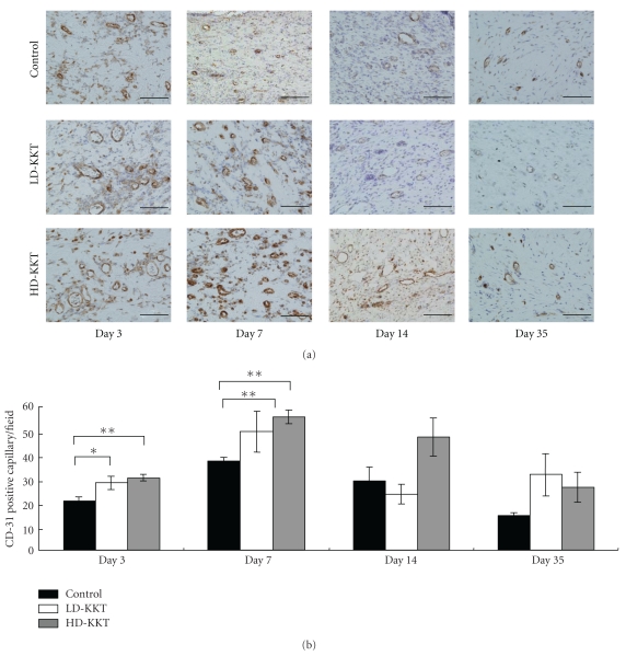
Figure 6.
CD-31 immunohistochemical findings of vessels at pressure ulcer edge on days 3, 7, 14, and 35. CD-31 immunopositive vessels are stained brown ((a); scale bar, 100 μm). The numbers of CD-31 immunopositive vessels per field (×200) were counted at wound tissue area including ulcer site, ulcer edge and granulation tissue (b). Values are expressed as mean ± SE (n = 4–7). *P < .05, **P < .01, versus control.

