
Figure 4.
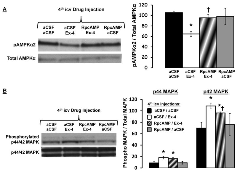
(A) 4th icv Ex-4 (0.3μg)-driven decrease phosphorylation in AMPKα2 in caudal DVC tissue lysates is PKA dependent, as 4th icv administration of the PKA inhibitor RpcAMP (20μg) attenuated the suppression in pAMPKα2 by Ex-4. Representative immunoblots for pAMPKα2 and total AMPKα are shown. * = P< 0.05 from aCSF/aCSF. † = P< 0.05 from aCSF/Ex-4. (B) RpcAMP administration attenuated the increased phosphorylation of p42-MAPK by Ex-4 administration, but did not alter the increased phosphorylation of p44-MAPK by Ex-4. Data are mean ± SEM. * = P< 0.05 from aCSF/aCSF. † = P< 0.05 from aCSF/Ex-4.