
Figure 5.
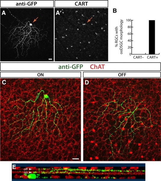
CART antibody labels ooDSGCs. A, CART and anti-GFP immunostaining identify double-positive RGCs in a retinal whole mount from line YFP-H. GFP channel shows the morphology of a YFP+ CART immunoreactive RGC (arrow; z-projection of confocal stack). CART channel (A′) shows CART+ RGCs in a single confocal plane through the GCL. Arrow indicates the soma of the YFP+CART+ double-positive cell. B, Morphological analysis of RGCs in line YFP-H that were CART+ and CART− (n = 140). All CART-immunoreactive RGCs (n = 22) have the bistratified morphology of ooDSGCs. By contrast, none of the CART− cells showed this morphology. C, D, Single confocal planes through the cell shown in A reveal morphological features that identify it as an ooDSGC. The cell dendrites (green) are bistratified, with both the ON (C) and OFF arbors (D) cofasciculating with the choline acetyltransferase (ChAT)-positive processes of starburst amacrines (red). E, Rotation of a 3-D reconstruction of the dendrites of this cell show that it has bistratified projections to the OFF and ON starburst IPL sublaminae (arowheads). GFP (green) and ChAT (red) channels are shown. Scale bars: 20 μm.