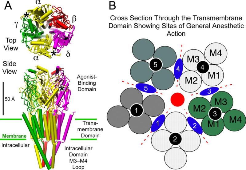
Figure 2.
General anesthetic binding sites on ligand-gated ion channels of the Cys-loop receptor superfamily. Panel A shows the structure of the nicotinic acetylcholine receptor (Unwin 2005), with its five subunits arranged centro-symmetrically around a central ion pore or channel, in both top view from the extracellular side and side view. The agonist site (*)in the extracellular agonist-binding domain is on the a-subunits in the interface with the g- and d-subunits. Panel B shows a schematic of a cross section through the transmembrane region. Each subunit, separated by dotted red lines, consists of four transmembrane helices shown as circles and numbered in the order they appear in the sequence. Three categories of anesthetic binding site (see text) are superimposed; intrasubunit sites (black circles); a channel lumen site (red circle), and subunit interface sites (blue lozenges). Molecular graphics images were produced using the UCSF Chimera package from the Resource for Biocomputing, Visualization, and Informatics at the University of California, San Francisco (supported by NIH P41 RR-01081) (74).

