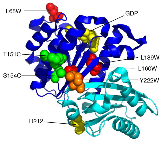
Figure 1.
Location of trp and cys mutants used in this study. The globular part of FtsZ comprises two subdomains (N-terminal dark blue, C-terminal cyan), which are independently folding (13, 14). GDP and the synergy residue D212 are shown in yellow spacefill. Three of the residues mutated to trp are shown in red spacefill. Y222 is in orange. The two residues mutated to cys (both in the presence of Y222W) are in green. The model is from the crystal structure of P. aeruginosa FtsZ, PDB 1ofu (39). The amino acids indicated are those of E. coli, some of which are different from the corresponding ones in P. aeruginosa.

