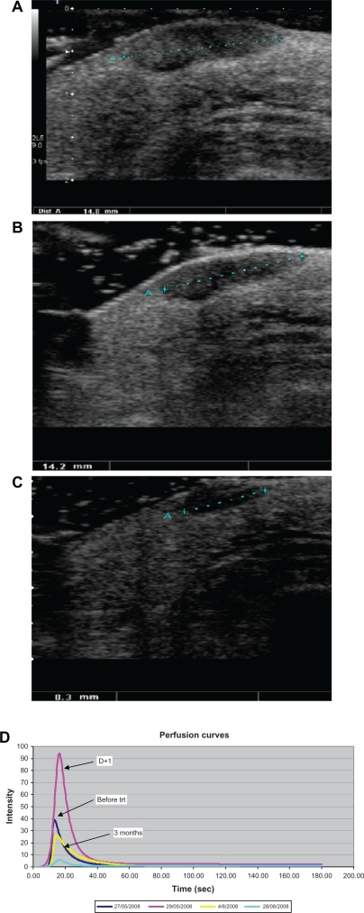
Figure 2.
In transit melanoma metastasis treated by isolated limb perfusion in a good responder. Ultrasound B mode (gray scale) scanning of superficial subcutaneous hypoechoic nodule of the right limb. A) Before treatment, the lesion was 14.8 mm in size. B) At day 1 after treatment no significant morphologic change was observed (14.2 mm). C) At day 7, the lesion shows a significant decrease in size (8.3 mm). D) In the same patient, after modelling, perfusion curves expressed as linear raw data show an important increase in perfusion parameters at day 1 after treatment compared with baseline.

