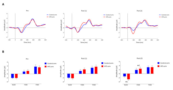
Figure 4.
Electrophysiological measurements. A) Grand average evoked potential waveforms. Plotted are the grand averaged evoked potentials waveforms for each measurement (pre, post (1) and post (2)) compared between the two arms (control vs. conditioned). Dotted line on X-axis represents stimulus onset. Upward is positive and downward is negative charge. B) Histograms representing the N100, P200 and P300 evoked potential amplitudes for each arm (control vs. conditioned) at every measurement (pre, post (1), post (2)). *1 The N100 amplitude observed at the conditioned arm was significantly (p < .05) larger than the N100 amplitude observed at the control arm 30 minutes after experimental conditioning stimulation (post (2)). *2 Averaged for both arms the P200 amplitude significantly (p < .05) increased between baseline (pre) and 30 minutes after experimental conditioning stimulation (post (2)).

