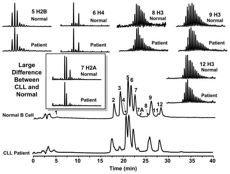
Figure 1.
LC-MS profile of core histones from a CLL patient and a normal volunteer. The relative level of two histone H2A variants in CLL cells was significantly different than that from normal B-cells. Additional changes in the abundance of two H3 variants (fractions 8 and 11) and two unidentified proteins (fractions 3 and 7A) were also observed.

