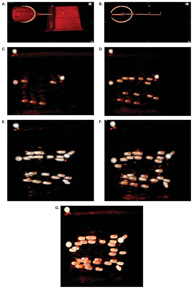
Figure 7.
Intra-operative verification of the placement of PPI seeds. Panels a and b show the robot head with the needle extended along with the phantom. The contrast window level has been modified in panel b compared to panel a to show the internal structure of the phantom. The three round white dots above the needle (easily seen in panel b) were glued to the outside surface of the phantom – they can be seen in each figure and serve as a reference point. Panel c, d, e, f, and g were taken after two, four, six, eight, and all ten needles, respectively, were placed. These figures show the ease of which the seeds can be identified in the CT environment. In contrast, reliable seed identification is nearly impossible using ultrasound imaging techniques.

