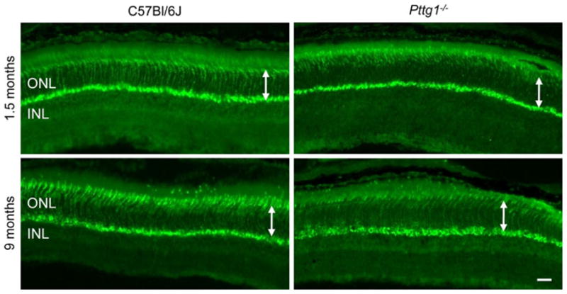
Fig. 4.

Mouse Cone Arrestin4 Immunohistochemical Staining in the Superior Region of Retinas from Pttg1−/− and C57Bl/6J Mice. IHC staining patterns with PAb mCAR-LUMIJ indicate a similar number of cone cells between Pttg1−/− mice and C57Bl/6J controls at both 1.5 and 9 months of age. White arrows demonstrate no change in ONL thickness. Scale bar, 10 μm
