
Figure 1.
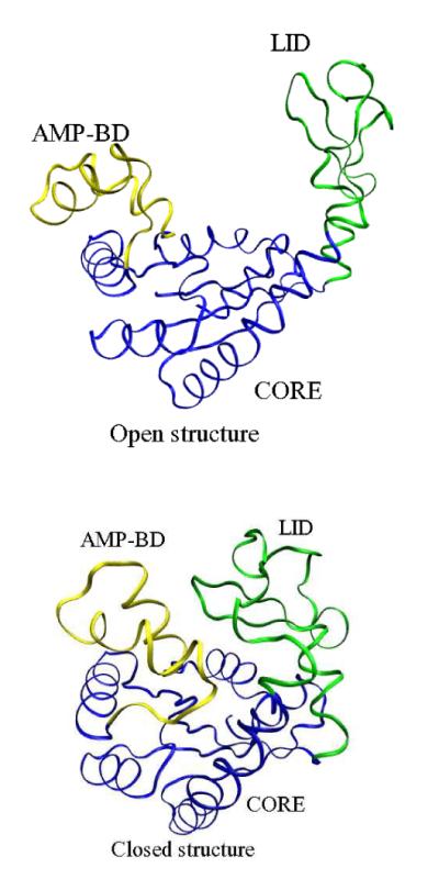
Crystal structures of adenylate kinase. The blue segment represents the core of the protein (CORE), the yellow segment is the AMP binding domain (BD), and the green segments is the flexible lid (LID). The left figure is the Apo (or Open) form, and the right figure is holo (Closed) form.