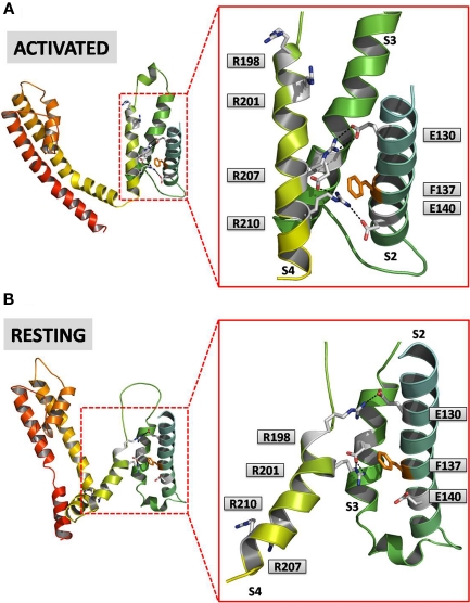
Figure 3.
An homology model for Kv7.2 channels. Both panels show side views of a single Kv7.2 subunit at lower (left) and higher (right) magnification. (A) Shows the activated configuration of the subunit and the corresponding VSD, whereas (B) shows the resting state. Amino acids involved in intra-subunit ion pairs in either resting and activated VSD configurations are shown. The homology model of the activated VSD of the Kv7.2 channel was generated as previously described (Miceli et al., 2008b); the resting state was derived from the corresponding model of the Kv1.2 channel described by Pathak et al. (2007).

