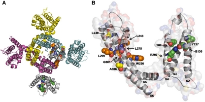
Figure 4.
An homology model for drug binding to Kv7.2 channels. Top view of the overall structure of the channel formed by four identical Kv7.2 subunits (A), and enlarged view of a single Kv7.2 subunit (B) captured in the activated configuration. The residues involved in binding of retigabine (indicated in orange), zinc pyrithione (indicated in yellow), and NH29 (indicated in green) highlighted. The L275 residue in the pore, common to both retigabine and zinc pyrithione binding, is indicated in violet. The S1 region has been removed for clarity. The homology model of the Kv7.2 channel was generated as previously described (Miceli et al., 2008b).

