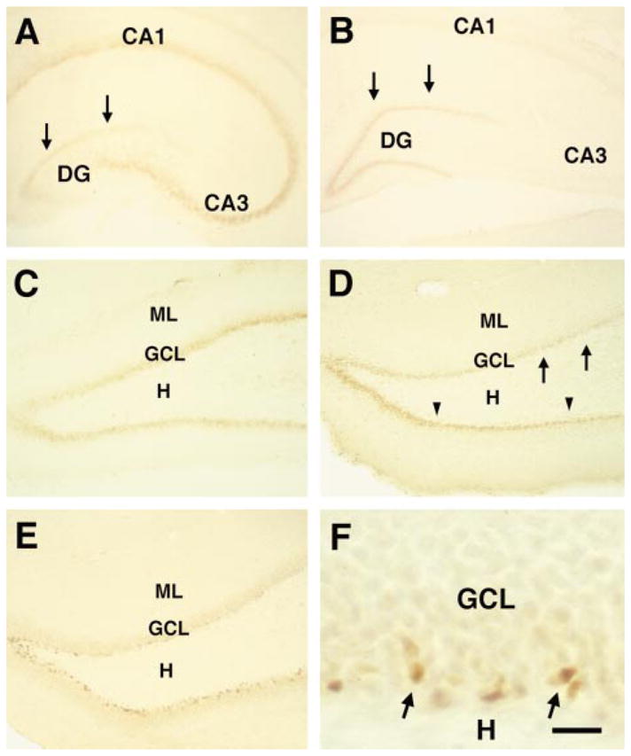
Fig. 1.

Activation pattern of CREB during postnatal hippocampal development. (A) In the hippocampal formation of the five day old rat (P5), phosphorylated CREB (pCREB) is clearly evident in the pyramidal layer (CA1 and CA3) compared with the granule cell (GC) layer (arrows). (B) By P12, hippocampal CREB activation pattern is radically altered; pCREB-immunoreactivity is almost undetectable in the pyramidal cell layer. In contrast, the GC layer (arrows), is now strongly immunoreactive. A higher magnification of the dentate gyrus (C) reveals that during the second postnatal week (P10) CREB is strongly phosphorylated in GCs, forming a 3–4 cells-deep, almost continuous band, along the hilar border of the GC layer. (D) By P18, pCREB-immunoreactive neurons still outline the hilar border, but in the earlier-maturing, lateral portion of the suprapyramidal blade (arrows), the density of immunoreactive cells is already diminished, whereas the hilar border of the infrapyramidal blade (arrowheads) remains intensely stained throughtout its length. (E) By P35, pCREB-immunoreactive neurons are scattered in a discontinuous, thin band at the GC layer hilar border. (F) a higher magnification of (E), shows variable, elongated or oval pCREB-labelled nuclei (typical of immature GCs), confined to the hilar border of the GC layer (arrows). CA, cornu ammonis; DG, dentate gyrus; GCL, granule cell layer; H, hilus; ML, molecular layer. Scale bar, 250 μm (A and B), 150 μm (C, D and E); 20 μm (F).
